Sign in to FlowVella

Forgot password?
Sign in with Facebook

New? Create your account

Sign up for FlowVella

Sign up with Facebook

Already have an account? Sign in now


By registering you are agreeing to our
Terms of Service

Share This Flow

Loading Flow

loading...

Downloading Image /

loading...

Downloading Image /

loading...

Downloading Image /

loading...

Downloading Image /

loading...

Downloading Image /

loading...

Downloading Image /

loading...

Downloading Image /

loading...

Downloading Image /

loading...

Downloading Image /

loading...

Downloading Image /

loading...

Downloading Image /

loading...

Downloading Image /

loading...

Downloading Image /

loading...

Downloading Image /

loading...

Downloading Image /

loading...

Downloading Image /

loading...

Downloading Image /

loading...

Downloading Image /

loading...

References & Resources

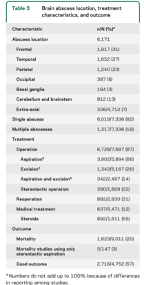
Brain Abscess

Clinical Pearls

Evaluation
Important:

- neuroimaging is the cornerstone of diagnosis
- false-negative rate of 6%
- preferential location: frontal and temporal lobes

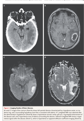
DWI superior to CT or conventional MRI in differentiating abscess from other lesions
- esp solitary tumours
- hyper intense on DWI and hypo intense on apparent diffusion coefficient images
- sens and spec of 96% (Xu, Clin Rad 2013)
PDF Preview

Downloading Image /

loading...

Downloading Image /

loading...
  • 1

  • 2

  • 3

  • 4

  • 5

  • 6

  • 7

  • 8

  • 9

  • 10

  • 11

  • 12

  • 13

  • 14

  • 15

  • 16

  • 17

  • 18

  • 19

  • 20